
- Diagnostics
Digital Motion X-Ray (DMX)
Digital Motion X-Ray (DMX) utilizes advanced diagnostic technology to detect and identify the ligament injuries that could never be seen before because of the lack of motion while weight bearing.
Position
Weight-bearing, standing
Ligaments Assessed
All 22 major cervical
Detects
Sub-failure injuries
Where
In office, same visit
Digital Motion X-ray (DMX) for Ligament Injury and Instability

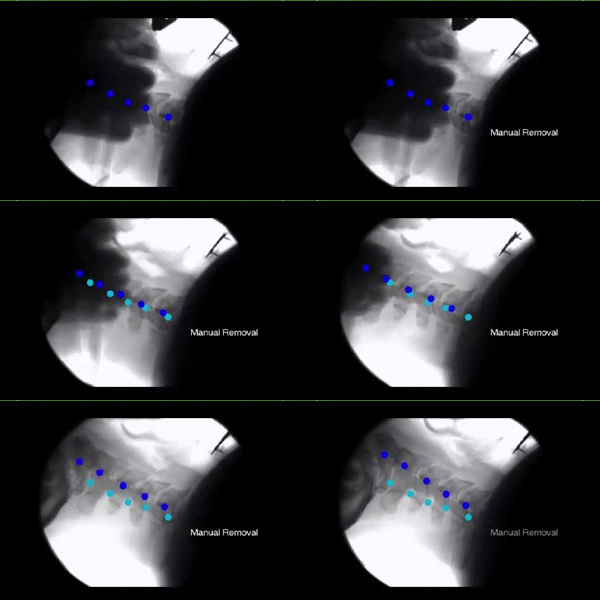
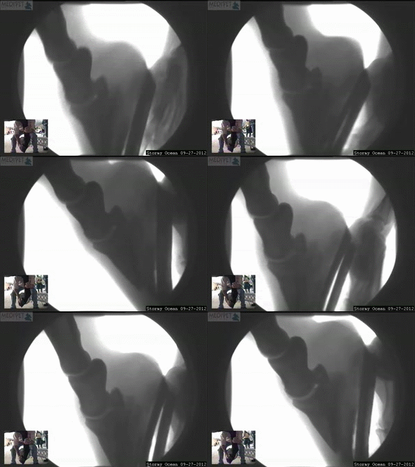
Real DMX study captures from our practice showing ligament motion analysis.
Static x-ray, MRIs and CTs often miss spinal ligament injuries because you don’t or can’t move during the study. The upper 30% of the cervical spine between the C0 (Occiput/Skull), C1 (Atlas), and C2 (Axis) posterior aspects of the spine contain no discs, just ligaments, and are not even evaluated on the majority of MRI studies.
Digital Motion X-ray (DMX) is the only diagnostic test with the ability to detect sub-failure ligament injuries. Motionless MRIs do not have the resolution power to detect the stretching or elongation of a cervical ligament, resulting in an inaccurate diagnosis. MRIs can identify a complete failure, more often seen in knees and shoulders — sub-failure ligament injuries are most commonly seen in the cervical spine, wrists, and TMJ.
DMX can provide valuable objective and measurable information about the stability of all 22 major ligaments in the cervical spine (anterior longitudinal ligament, posterior longitudinal ligament, interspinous ligaments, capsular ligaments, alar ligaments, transverse ligament, and accessory ligaments). It is often utilized to identify and demonstrate post traumatic ligament instability in the neck that may be responsible for neck pain, dizziness, headaches, unsteadiness, and referred pain to the shoulders, arms, and/or hands — symptoms caused by stretched ligament injuries which cannot be detected by MRI.
DMX testing is performed with the patient in a weight bearing standing position, moving the cervical spine through different motions. Each motion tests the function of specific groups of cervical ligaments and is an independent study focused on the anatomical structures found in that motion. Ligamentous injuries are painful, progressive, and permanent, so they need to be analyzed and detected for a proper diagnosis and to determine the severity of the injury.
American Medical Association (AMA) Guidelines for Permanent Impairment (Fifth Edition) rate ligamentous injuries for impairment. If one or more ligaments are damaged, 5–8% whole body permanent impairment can be assigned; if aberrant motion exceeds 3.5mm, 25% whole body permanent impairment can be assigned. Damaged ligaments heal by fibrous repair (scar tissue), not by regeneration of the original elastin tissue — leaving the injured ligament permanently weaker and more susceptible to future injury. The altered biomechanics of an unstable joint lead to progressive degenerative changes 6–10 times faster than normal.
Anyone who has been in car accidents (even a minor one), or has had traumatic falls or injuries, can benefit from the DMX digital motion X-ray study and computerized measurements.
Digital Motion X-Ray
Unlike traditional X-rays, which only capture still images, Digital Motion X-Ray allows us to observe real-time movement, providing a more comprehensive view of how your joints, ligaments, and muscles function during motion. This motion-based imaging is essential for diagnosing conditions like ligament tears, instability in the spine, joint dysfunction, and more.
One of the standout features is its ability to identify subtle abnormalities that are often missed in conventional X-rays — ligament instability and joint laxity in particular, conditions that can lead to chronic pain, discomfort, and limited mobility.
For athletes, DMX is a valuable tool for diagnosing joint and ligament injuries caused by high-impact activities — sprained ankles, torn ACLs, or shoulder injuries. For those suffering from chronic pain or unexplained symptoms, DMX can uncover issues that might otherwise go unnoticed, such as joint misalignment, instability, or inflammation.
DMX is also essential for evaluating spinal issues, particularly spinal instability and degenerative disc disease, revealing how vertebrae interact and whether there is abnormal motion contributing to pain. The procedure itself is non-invasive: the patient moves in specific ways while the system captures high-quality images, with results available almost immediately.
[The full disabled source contains two near-verbatim repetitions of this expanded article — condensed here to preserve its substance without needless duplication. It links to the National Institute of Biomedical Imaging and Bioengineering’s page on X-rays.]
Schedule a DMX Study
Most Major Insurances Accepted. Call for more information or book an apt online.
- Office hours
- Monday: 9am to 7pm
- Tuesday: 3pm to 7pm
- Wednesday: 9am to 7pm
- Thursday: 10am to 7pm
- Friday–Sunday: Closed
- Who it's for
Reasons We Order DMX
Whiplash from an auto accident, even a minor one
Traumatic falls or injuries
Neck pain, dizziness or headaches with a normal MRI
Unsteadiness or referred pain to the shoulders, arms or hands
Suspected TMJ or wrist ligament instability
Documentation for AMA permanent impairment ratings
Legal or workers' compensation case documentation
Pain that doesn't match static imaging findings
- What to expect
The Visit, Step by Step
1
Evaluation First
2
Weight-Bearing Study
3
Computerized Measurement
4
Review & Report
- Our promise
Every patient receives a careful and thorough evaluation before any treatment plans are made. This highly individualized level of care is critical to make the correct diagnosis and pick the best, most effective course of treatment for each patient. This level of care is beyond most busy clinics that treat patients by standard protocols only.
Objective documentation
Measurable evidence of ligament instability that static imaging cannot capture.
A specific diagnosis
The involved ligament group is identified, not guessed at.
Impairment rating support
Findings can support AMA Guidelines permanent impairment ratings when applicable.
An early baseline
Catching instability early may help avoid faster degenerative change down the line.
Related Treatments
Electrodiagnostic Testing
Non-Surgical Spinal Decompression
Discover the Freedom of a Pain-Free Life!
Most Major Insurances Accepted. We do not participate in Medicaid plans.
- Visit us
20433 Bruce B Downs Blvd, Tampa, FL 33647
Office Hours
- Monday: 9am to 7pm
- Tuesday: 3pm to 7pm
- Wednesday: 9am to 7pm
- Thursday: 10am to 7pm
- Friday–Sunday: Closed